
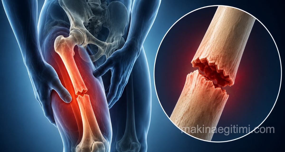
The femur is the longest and strongest bone in the human body; a fracture of it is a serious condition and, in adults, almost always requires surgery. In young people it results from high-energy trauma such as traffic accidents, while in the elderly it typically results from a simple fall linked to bone loss. Bone healing generally takes 6–12 weeks; what truly determines the outcome is early movement after surgery and consistent rehabilitation.
The thigh bone (femur) is the longest, thickest, and strongest bone in our body. Breaking such a solid bone normally requires a large force — a traffic accident, a fall from height.
There is, however, one exception: in an elderly person with bone loss, even tripping on a rug at home can lead to a femur fracture.And this group makes up the most common — and highest-risk — segment of all femur fractures.
In this article we explain how femur fractures occur, treatment methods, a realistic recovery timeline, and — perhaps most importantly — what can be done at home to prevent this fracture in elderly individuals.
What is a femur fracture?
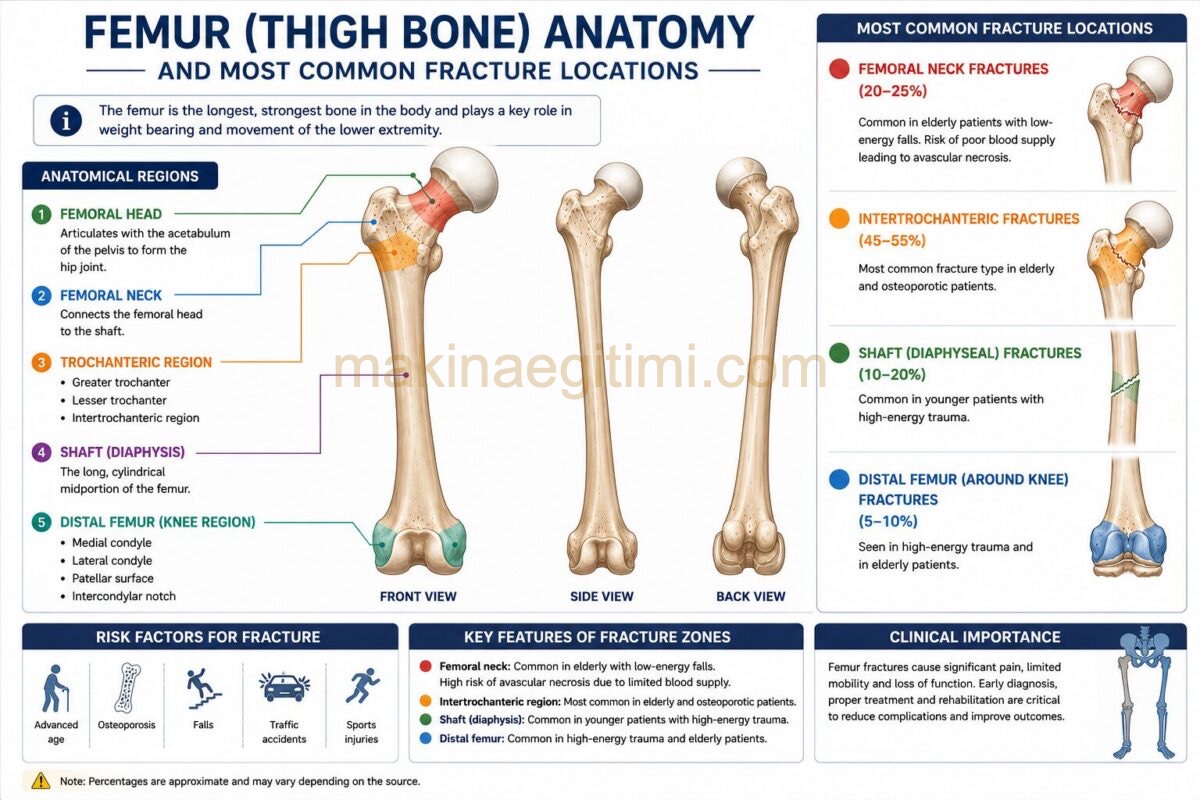
The femur is the thigh bone that runs between the hip joint and the knee joint. Its upper end is ball-shaped and sits in the socket of the pelvic bone to form the hip joint; its lower end forms part of the knee joint.
Which region the fracture is in completely changes the treatment and the recovery:
| Fracture region | Most common in | Characteristic |
|---|---|---|
| Femoral neck (hip fracture) | Elderly people, those with bone loss | Blood supply to this region is delicate; healing problems can occur |
| Trochanteric region (hip fracture) | Elderly people | Blood supply is good, chance of healing is high |
| Femoral shaft (thigh fracture) | Young people, after an accident | Requires great force, can cause serious bleeding |
| Knee end | Any age | Involves the knee joint, carries a risk of limited motion |
What is commonly called a “hip fracture” actually refers to fractures in the upper part of the femur.
Why does a femur fracture occur?
Femur fractures occur in two different patient groups, for two different reasons:
1. In the elderly: bone loss + a fall
This is the most common scenario. As we age, bone density decreases (osteoporosis) and the bone becomes fragile. In this situation, even a simple fall that would cause no harm to a healthy bone can lead to a fracture.
Factors that increase the risk:
- Bone loss (osteoporosis)
- The postmenopausal period in women — declining estrogen accelerates bone loss
- Balance problems, dizziness, difficulty walking
- Vision problems
- Medications that can cause dizziness, such as sleeping pills and blood pressure medication
- Vitamin D and calcium deficiency
- Loss of muscle mass
- A home environment that creates a fall risk: loose rugs, cables, slippery bathroom floors, insufficient lighting
2. In young people: high-energy trauma
- Traffic accidents (especially motorcycle accidents)
- Falls from height
- Severe sports injuries
- Workplace accidents
3. Rarer causes
- Fatigue (stress) fractures: Cracks that develop from repetitive strain in long-distance runners and military personnel.
- Pathological fractures: Fractures that occur with very minor strain when a disease weakening the bone (tumor, cyst, infection) is present.
What are the symptoms of a femur fracture?
In most cases the symptoms are quite clear and severe:
- Severe hip or thigh pain
- Inability to stand up or bear weight on the leg
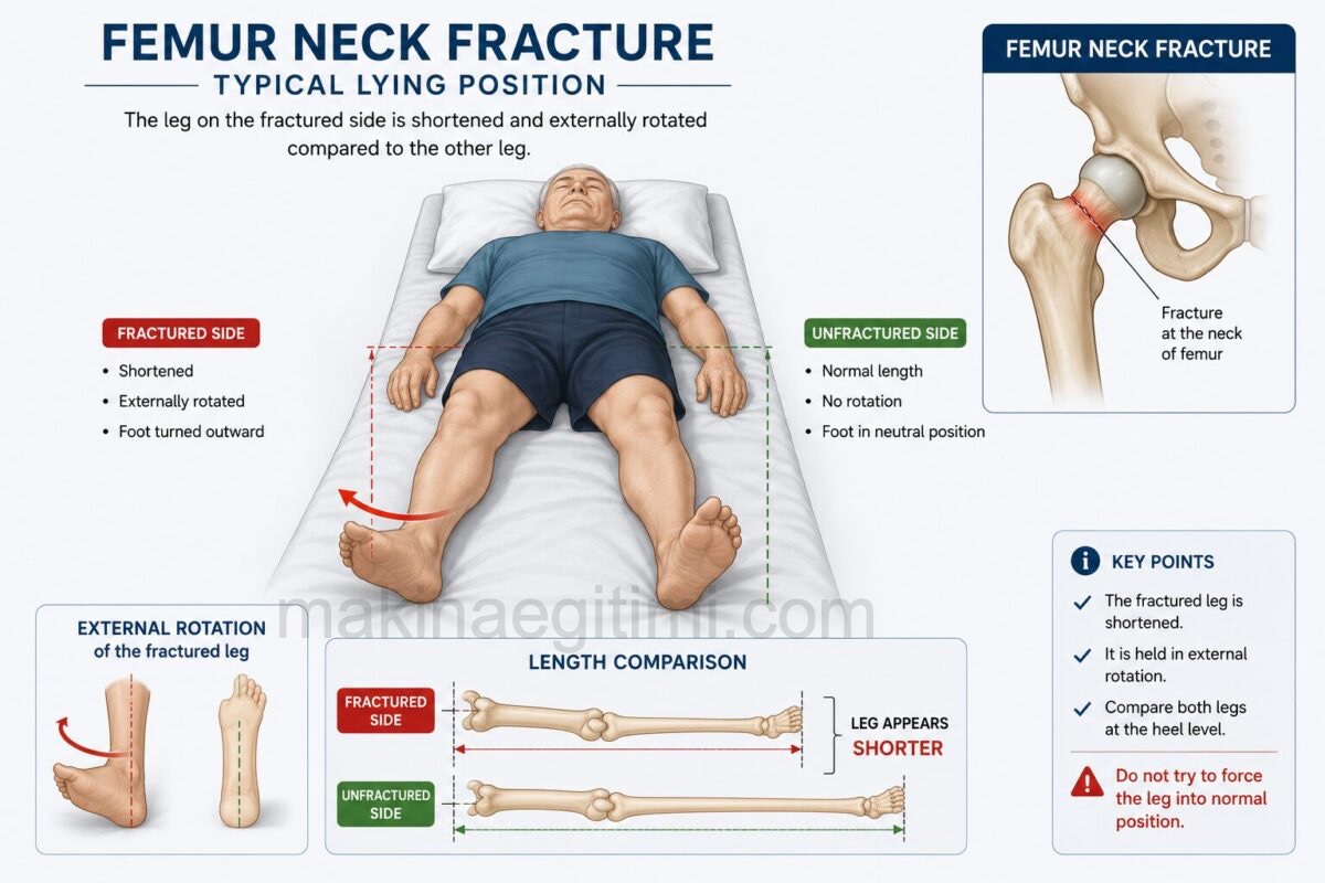
- The leg turning outward
- The fractured leg appearing shorter than the other
- Unbearable pain when the hip is moved
- Swelling and bruising in the thigh area
- In femoral shaft fractures, a visible deformity in the thigh
⚠️ An important exception to be aware of: A femur fracture is not always this dramatic. Especially in non-displaced cracks related to bone loss, a person may be able to stand, or even walk with a limp. The only complaint may be pain in the groin area. For this reason, if an elderly person has persistent groin or hip pain after a fall, an X-ray should be taken even if they are able to walk.
Is a femur fracture contagious?
No. A fracture is a disruption of the bone’s integrity caused by trauma; it is not caused by a pathogen and cannot be contagious.
However, the underlying bone loss is a different matter: bone loss is also not contagious, but it does have a hereditary predisposition. A woman whose mother had a history of hip fracture has an increased risk of fracture herself. For this reason, people with a family history of hip fracture are advised to have a bone density measurement.
How is a femur fracture diagnosed? Which department should I go to?
Where you should go: directly to the Emergency Department. An appointment should not be waited for if a femur fracture is suspected. Treatment is carried out by an Orthopedics and Traumatology specialist.
Examinations performed:
- Physical examination: The position of the leg, length discrepancy, and limitation of movement are assessed.
- X-ray: The basis of diagnosis; it clearly shows most fractures.
- CT or MRI: Needed when a non-displaced crack not visible on X-ray is suspected. In elderly patients, these tests are ordered if pain continues even when the X-ray is normal.
- Blood tests and general assessment: Evaluation of heart, lung, and kidney status before surgery.
First aid: if this happens near you
- Absolutely do not try to move the person or get them to stand up.
- Do not try to straighten the leg.
- Call emergency services.
- Keep the person warm and calm.
- Since surgery may be needed, do not give them anything to eat or drink.
- If there is an open wound, cover it with a clean cloth without pressing on it.
How is a femur fracture treated?
In adults, almost all femur fractures are treated with surgery. The reason isn’t just to get the bone to heal properly — the real goal is to get the patient back on their feet as soon as possible.
Why is surgery so important?
This may be the most critical piece of information in this article. Especially for an elderly patient, staying in bed for weeks is more dangerous than the fracture itself. Prolonged bed rest leads to:
- Lung infection (pneumonia)
- Blood clot formation in the leg veins and that clot traveling to the lungs
- Pressure sores
- Rapid muscle loss — every week spent in bed causes significant loss of muscle mass
- Urinary tract infections
- Worsening of existing heart, diabetes, and kidney conditions
- Mental decline and depression
For this reason, the goal in orthopedic practice is to take the patient to surgery within the first 24–48 hours as much as the patient’s overall condition allows. Delaying surgery significantly increases the risk of complications.
Surgical methods
Which method is used is determined by the location and type of the fracture, the patient’s age, and bone quality:
1. Fixation with a nail (intramedullary nail). A titanium nail placed into the bone’s internal canal supports the fracture from within. This is the most commonly used method for femoral shaft and trochanteric hip fractures. Its key advantage: most patients can be gotten up and can bear weight on the leg the day after surgery.
2. Fixation with a plate and screws. A metal plate placed on the outside of the bone is secured with screws. With this method, full weight-bearing is generally not allowed for about 6 weeks.
3. Cannulated screws. Preferred in young patients and in non-displaced femoral neck fractures, in order to preserve the patient’s own bone.
4. Partial (hemi-) arthroplasty. In femoral neck fractures, the head of the bone is removed and replaced with a prosthesis. Generally preferred in older patients with additional health conditions who walk short distances. Its advantage: the patient can usually bear weight and walk right away.
5. Total hip replacement. Both the femoral head and the hip socket are replaced. Considered for more active patients in good general health.
6. External fixation. Used as a temporary solution in severe open fractures or when the patient’s condition is not suitable for surgery.
When is non-surgical treatment considered?
Only in very limited situations: in small, non-displaced cracks, or in patients for whom the surgical risk is high enough to be life-threatening. Because this approach requires prolonged immobility, the risk of complications is high, and it is an exceptional choice.
The recovery process: a realistic timeline
This is what patients and their families want to know most. Timelines vary by individual, fracture type, and age; the table below is a general framework:
| Period | What happens |
|---|---|
| Day 1 after surgery | Movement in bed with a physiotherapist, ankle exercises, standing with support for most patients |
| First week | Discharge from hospital, walking training with a walker or crutches |
| Week 2 | Wound-healing check, removal of stitches |
| Week 6 | First check of bone healing; if a plate was used, weight-bearing generally begins around this time |
| Weeks 6–12 | Bone healing largely complete |
| Months 3–6 | Regaining muscle strength and walking quality |
| Months 6–12 | Full recovery; return to sports in some patients |
Factors that slow healing: Smoking (the most important), diabetes, bone loss, poor nutrition, advanced age, comminuted (fragmented) fracture.
Rehabilitation: the true determinant of recovery
Surgery is only half the job. The other half is physical therapy, and if it is neglected, even a successful surgery cannot get the patient walking again.
A rehabilitation program generally includes:
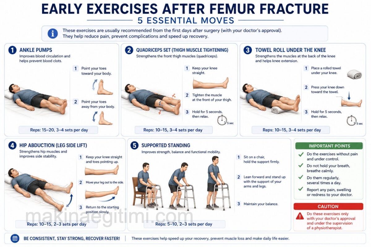
- Ankle pumping exercises — from the first day, to prevent clot formation
- Knee and hip range-of-motion exercises — to prevent joint stiffness
- Muscle strengthening — especially the front thigh muscles and hip muscles
- Walking training — progressing from a walker to crutches, then to a cane
- Balance work — critically important to prevent a new fall
- Stair climbing training
⚠️ See a doctor immediately after surgery if you notice these symptoms
- One-sided swelling, redness, warmth, and pain in the calf — may indicate a blood clot in the leg vein
- Sudden shortness of breath, chest pain, coughing up blood — may indicate the clot has traveled to the lungs, this is an emergency
- Fever, chills, discharge or foul odor from the wound — sign of infection
- Increasing pain at the surgical site and a new deformity in the leg
- New numbness, tingling, or color change in the leg
- Confusion, excessive drowsiness, new-onset mental disorientation — especially important in the elderly
What should you pay attention to during treatment?
Move early. The “let the fracture heal first, then I’ll get up” approach is wrong and dangerous. Move as much as your doctor allows.
Follow the weight-bearing instructions to the letter. Depending on which method was used, you will be told “you can bear full weight,” “you can bear partial weight,” or “you must not bear weight at all.” Exceeding this instruction can cause the fracture to shift out of place.
Don’t skip rehabilitation. Stopping physical therapy is the most costly mistake in this process.
Take precautions against bedsores: There are air mattresses, also known as air mattresses, that are placed under the patient’s sheets and inflate and deflate sections using an electric motor. They are recommended for patients who have to lie down for long periods. The electric motor of these products operates quietly so as not to disturb the patient’s sleep.

Also, periodically check the areas of the patient’s body that come into contact with the bed. Consult your doctor if you notice any unusual redness.
If you’ve had a prosthesis fitted, pay attention to movement restrictions. In the early period, bending the leg too far toward the body, crossing your legs, and turning the leg inward can cause the prosthesis to dislocate. Avoid sitting in low chairs and using low toilets; use a raised toilet seat.
Take precautions against constipation. Inactivity and pain medication cause constipation. Drink plenty of fluids and eat fiber-rich foods.
Quit smoking. It directly delays bone healing; quitting is the habit change that makes the biggest difference in fracture recovery.
Don’t neglect osteoporosis treatment. This is the most commonly skipped step. An elderly patient who has had one fracture going on to break their other hip a year later is a common, and preventable, occurrence in orthopedics. A first fracture is a strong warning sign that the skeletal system has weakened. Get a bone density measurement (DXA) before discharge or at follow-up visits, and start osteoporosis treatment if needed.
Make your home safe: a fall-prevention checklist
This section covers the most practical steps for preventing a new fracture:
- Remove loose rugs and mats, or secure them with non-slip tape
- Gather floor cables against the wall
- Install grab bars in the bathroom and toilet
- Use a non-slip mat in the shower, and a shower chair if possible
- Add a night light; especially light the path between the bedroom and the bathroom
- Place frequently used items on shelves you can reach without stretching
- Wear closed-back, non-slip shoes instead of slippers
- Have handrails installed on both sides of staircases
- Have your eyeglass prescription checked once a year
- Review your medications with your doctor — sleeping pills, blood pressure medication, and some psychiatric medications can cause dizziness and increase fall risk
- If you have a pet, be careful of it getting underfoot
Nutrition, herbal supplements, and lifestyle: what can help?
Nutrition plays a bigger role in fracture healing than most people assume. As the body rebuilds bone tissue, it needs both building blocks and energy.
Foods that may help
- Protein — the most important nutrient. About half of bone tissue is made up of protein, and protein needs increase during fracture healing. It also prevents muscle loss after surgery. Eggs, milk and dairy products, meat, chicken, fish, legumes. Inadequate protein intake in elderly patients is the most common nutritional problem that delays healing.
- Calcium sources: Milk, yogurt, cheese, kefir, sesame, dark leafy greens, small fish eaten with their bones.
- Vitamin D: Essential for calcium to be absorbed from the intestine. Egg yolk, oily fish, and most importantly sunlight. Vitamin D deficiency is very common in our country; be sure to have your level checked.
- Vitamin C: Plays a role in forming the collagen structure in bone. Citrus fruits, red pepper, rosehip, parsley.
- Zinc and magnesium: Pumpkin seeds, almonds, red meat, whole grains.
- Vitamin K: Leafy green vegetables.
- Adequate fluids and fiber-rich foods: Prevents constipation during the period of inactivity.
Things to limit
- Smoking — the factor that delays bone healing the most
- Excessive alcohol — impairs bone formation and increases fall risk
- Excessive caffeine — can increase calcium loss when consumed in very high amounts
- Excessive salt and processed foods
An honest note about herbal supplements
You often hear about herbal mixtures and ointments claimed to speed up fracture healing. Here’s what you should know about them:
- None of them change how long a fracture takes to heal. Bone heals on its own biological timetable.
- If you are taking blood-thinning medication after surgery (often given to prevent clots), most herbal products increase bleeding risk. Ginkgo biloba, high-dose turmeric, and garlic supplements are at the top of this list.
- Mixtures applied externally to the fracture site carry a risk of skin irritation and wound infection. Do not apply anything to the surgical wound.
- Tell your doctor about every supplement you want to use.
What helps most in the long run
Maintaining muscle strength and balance. The only effective way to increase bone density and prevent falls is regular, weight-bearing exercise: brisk walking, light resistance work, balance exercises. Balance training such as tai chi, done under expert guidance, has been reported to reduce falls in the elderly.
Frequently asked questions about femur fractures
Bone healing generally takes 6–12 weeks, though in some cases this can extend to a year. Regaining muscle strength and walking quality takes 3–6 months. Smoking, diabetes, and bone loss are the main factors that delay healing.
In most patients, standing with support, guided by a physiotherapist, begins the day after surgery. If the fracture was fixed with a nail, weight can usually be put on the leg the following day; if fixed with a plate, full weight-bearing generally begins after 6 weeks. This decision belongs entirely to your doctor.
The danger comes less from the fracture itself and more from the complications caused by inactivity: lung infection, blood clots in the leg veins, pressure sores, and rapid muscle loss. For this reason, the goal is to operate as early as the patient’s overall condition allows and get them back on their feet quickly.
In adults, surgery is required except in very limited situations. Non-surgical follow-up is only considered for small, non-displaced cracks or in patients for whom the surgical risk is high enough to be life-threatening; because this approach requires prolonged immobility, its complication risk is high.
If a femur fracture is suspected, you should go directly to the Emergency Department; an appointment should not be waited for. Treatment is carried out by an Orthopedics and Traumatology specialist. In elderly patients, an evaluation by Internal Medicine or Endocrinology for bone loss may also be needed.
Related videos you can watch
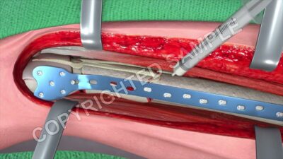
Femur Fracture Open Reduction and Internal Fixation| ORIF
Hip Fracture – Treatment, Recovery & Prevention Tips
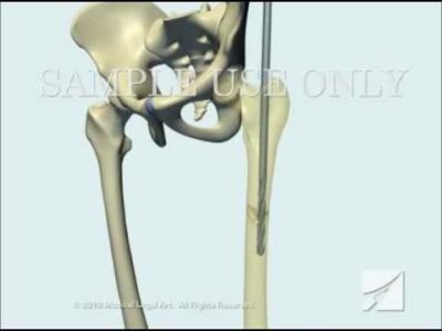
Femoral Fracture Fixation Surgery: Intramedullary Nail
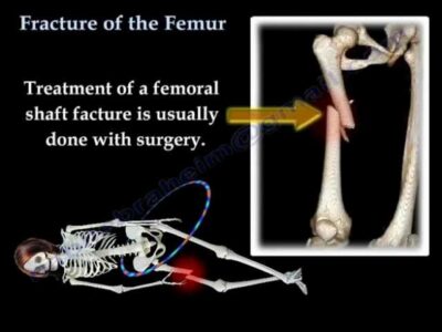
Fracture of the Femur and its fixation – Everything You Need To Know – Dr. Nabil Ebraheim
Medical disclaimer: This article is for general informational purposes only; it does not replace an examination, diagnosis, or treatment. Be sure to consult a doctor for your symptoms. Do not stop or change any medication without consulting your doctor.
Related Posts
 What Is Type 2 Diabetes? Symptoms and Treatment
 What Is a Herniated Lumbar Disc? Symptoms, Causes and Treatment
 Cataract: Symptoms, Causes and Surgery
 Rheumatoid Arthritis: What Is It? Symptoms and Treatment
 What Is a Herniated Cervical Disc? Symptoms, Causes and Treatment
 What Is Arteriosclerosis? Symptoms, Causes, and Treatment
 High Blood Pressure (Hypertension): Causes, Symptoms and Treatment
 What Is Reflux? Symptoms, Causes, and Treatment Methods