
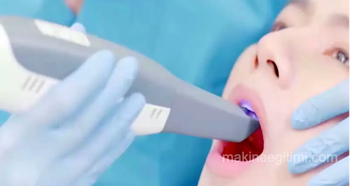
The Best Intraoral 3D Scanners for Faster Dental Impressions. Intraoral scanning is rapidly replacing molding for accurate measurements in digital dentistry.
For decades, the only way to obtain a clean dental impression for making dentures, crowns, implants, or aligners was to bite down on a large tray of plaster or acrylic molding adhesive.
Nowadays, intraoral scanners are becoming more popular in dentistry for a variety of reasons. These scanning wands offer a relatively easy and patient-friendly way to create optical impressions of the inside of a person’s mouth.
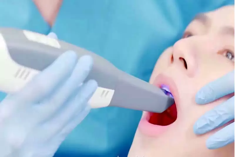
These handheld devices rapidly produce a full-color 3D model of a patient’s teeth and gums. The 1:1 digital copy can then be used to diagnose abnormalities, assist with surgery, and create precise prosthetic devices.
3D scans can be viewed instantly on a computer or touchscreen for better communication and collaboration between the patient and the practitioner.
How Do Intraoral Scanners Work?
A scanning wand, about twice the size of an electric toothbrush, is inserted into the patient’s mouth to collect visual data.

The dentist moves it slowly to capture an impression of the teeth and gums from as many angles as possible.
Unlike a plaster mold, which can only be placed once, a scanner can repeatedly scan an area until a complete rendering of the entire arch is finished.
Scanners work by projecting a light source, such as a laser (without heat or harmful effects), or structured light, onto the patient’s dental arches. The images are then processed by scanning software that takes the data points generated by the device and combines them to create a “point cloud”. The software then triangulates the point clouds using mathematical models that analyze the data and produce a final, precise 3D visualization of the dentogingival tissues.
Scanner Software Is Key
Once the intraoral scanner software converts the captured images into digital models, these images can then be sent to in-house or external labs that design and manufacture a prosthesis. In some cases, all of this — including 3D-printing the prosthesis — can happen chairside. If the scan is for diagnostic purposes only, the dentist can show the patient the problem areas and use the image to develop a treatment or surgical plan.

Software accuracy is key so that the maximum amount of data from the scans can be combined and stored. That’s why many intraoral scanners come with their own software for converting captured images into these digital models.
These devices have obvious advantages, including comfort, time efficiency, and better communication between practitioners, patients, and labs. However, the devices are expensive, starting at around $12,000.
Ultimately, patients will likely prefer the relative comfort of intraoral scanners, and will be able to communicate more effectively with dentists if they can see an image of their dental issues. Combined with the elimination of the radiation exposure used by some earlier imaging techniques, and the time saved by being able to scan chairside in real time, the cost-benefit analysis will favor these devices for most dental practices.
Digital Impression Taking
Intraoral scanners collect tooth and gum data digitally, instead of a traditional plaster impression.
Patient Comfort
Digital scanning is a much more comfortable and faster process for patients compared to traditional impression materials.
CAD/CAM Integration
Intraoral scanner data can be transferred directly into CAD/CAM systems for use in manufacturing prosthetics and crowns.
Related Questions
In the traditional method, the patient has to hold a sticky substance in their mouth for a long time as it hardens, which is uncomfortable. Since an intraoral scanner only performs an optical scan, this discomfort is avoided entirely.
The patient can view a 3D model of their own mouth live, together with the dentist. This lets the dentist explain the situation to the patient concretely, and allows the treatment decision to be made together, with more informed input.
Related Posts
